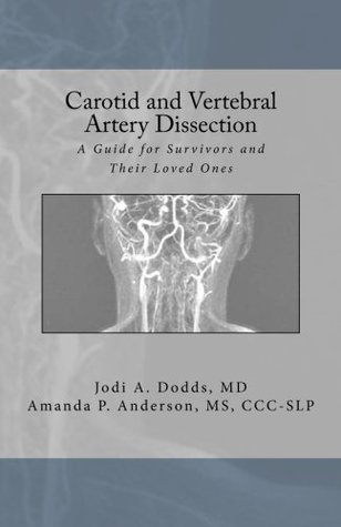
Read Carotid and Vertebral Artery Dissection: A Guide For Survivors and Their Loved Ones - Jodi A Dodds MD file in ePub
Related searches:
Carotid Artery Disease: Causes, Risk Factors, and Treatment
Carotid and Vertebral Artery Dissection: A Guide For Survivors and Their Loved Ones
Common Carotid Artery: Anatomy, Function, and Significance
Carotid and vertebral artery dissection syndromes
Carotid, Vertebral and Intracranial Artery Stenosis
A Spectrum of Doppler Waveforms in the Carotid and Vertebral
[Full text] Incidence of traumatic carotid and vertebral artery
Blood flow in internal carotid and vertebral arteries during orthostatic
Spontaneous Dissection of the Carotid and Vertebral Arteries NEJM
SPONTANEOUS DISSECTION OF THE CAROTID AND
Imaging in Carotid and Vertebral Artery Dissection: Practice
Critical carotid and vertebral arterial occlusive - AHA Journals
Dissection of the carotid and the vertebral artery - AMBOSS
Headache and neck pain in spontaneous internal carotid and
Carotid artery disease - Symptoms and causes - Mayo Clinic
Ch. 20 Key Terms - Anatomy and Physiology OpenStax
Carotid Duplex: Purpose, Procedure, and Preparation
Carotid and Vertebral Artery Doppler Ultrasound Waveforms: A
Major Arteries of the Head and Neck - Carotid - TeachMeAnatomy
Carotid and Vertebral Artery Insufficiency - JAMA Network
Collaterals between the External Carotid Artery and the Vertebral
Aberrant Origin of Vertebral Artery and its Clinical Implications
Carotid and Vertebral Arteries Radiology Key
[Clinical characteristics of internal carotid and vertebral
Vascular resistance of carotid and vertebral arteries is
(PDF) Carotid and vertebral artery dissection syndromes
Extracranial Carotid and Vertebral Artery Disease
Carotid and Vertebral Artery Intervention Thoracic Key
Abnormalities of the head and neck arteries (Cerebrovascular
Carotid and vertebral artery dissections: three-dimensional
CAROTID AND VERTEBRAL ARTERY DISSECTIONS - ScienceDirect
Bilateral Carotid and Vertebral Artery Dissection from Blunt
Editor's Choice - Management of Atherosclerotic Carotid and
Surgical Anatomy of Carotid and Vertebral Arteries SpringerLink
Extracranial Carotid and Vertebral Arteries Thoracic Key
MRA of the Carotid and Vertebral Arteries - W-Radiology
REVIEW Carotid and vertebral artery dissection syndromes
Extracranial carotid and vertebral artery disease - Oxford
Vertebral Artery Segments, Stenosis and Artery Dissection
Blood-Flow Volume Quantification in Internal Carotid and
Editor’s choice — management of atherosclerotic carotid and vertebral artery disease: 2017 clinical practice guidelines of the european society for vascular surgery (esvs).
Transient compression of the vertebral arteries can also cause injury, dissection of the artery, more permanent blockage of the artery, and transient mini-stroke.
Carotid artery disease occurs when fatty deposits (plaques) clog the blood vessels that deliver blood to your brain and head (carotid arteries). The blockage increases your risk of stroke, a medical emergency that occurs when the blood supply to the brain is interrupted or seriously reduced.
The carotid arteries supply blood to the large, front part of the brain, where thinking, speech, personality and sensory and motor functions reside. The vertebral arteries run through the spine and supply blood to the back part of the brain (the brainstem and cerebellum).
The vertebral artery, a component of the vertebrobasilar artery system, supplies 20% of the blood to the brain (primarily the posterior cranial fossa), with the remaining 80% being supplied by the carotid system. The vertebral artery supply blood to the brainstem, spinal cord, and to the vertebrae and their associated ligaments and muscles.
Carotid and vertebral arteries lie on either side of the neck(1). Carotid arteries consist of three layers: tunica intima (the inner layer), tunica media (the middle layer), and tunica adventitia (the outer layer(2).
Cpsd runs several research studies looking into the causes, investigation, and management of large artery atherosclerosis, carotid stenosis, vertebral artery.
Carotid artery stenosis is a disease of the arteries that carry blood to the brain. Stenosis leads to progressive narrowing of the artery and eventual restriction of blood flow to this vital organ.
Carotid and vertebral artery disease affects a large segment of the population with the potential of causing severe disability from a major stroke. This book places emphasis on the medical, endovascular and surgical approaches in managing patients with extracranial carotid and vertebral artery disease following pertinent diagnostic studies.
The buildup of cholesterol plaques in your carotid arteries can create blood clots.
What is carotid artery disease? your carotid arteries are the major blood vessels that deliver blood to your brain.
Dissections involving the carotid and vertebral arteries, once believed to be rare, have been reported with increasing frequency as a cause of spontaneous and post-traumatic stroke in young adults. 2, 9 20, 50 recognition of cervical arterial dissection, however, is not always straightforward.
The carotid arteries are blood vessels that supply blood to the head, neck and brain. Sebastian kaulitzki / science photo library / getty images arteries are vessels that carry blood away from the heart.
17 jul 2018 as a result, direct visualization of the intramural hematoma, simultaneous examination of the carotid and vertebral arteries, precise determination.
22 mar 2001 described dissections of carotid and vertebral arteries as detected by modern diagnostic approaches, that dissections began to be routinely.
The head and neck receives the majority of its blood supply through the carotid and vertebral arteries.
Objective: to compare demographic, clinical, and imaging characteristics of patients with internal carotid artery dissection (icad) and vertebral artery dissection (vad) in a russian population.
3 nov 2020 cervical artery dissections is the collective term for dissections of the carotid or vertebral arteries they are important causes of stroke in younger.
Guideline on the management of patients with extracranial carotid and vertebral artery disease.
13 jan 2021 the vascular supply to the brain is divided into the anterior and posterior circulations originating from the carotid and vertebral arteries,.
Doppler ultrasound is the most cost-effective imaging modality for screening severe carotid artery atherosclerotic disease.
In carotid artery dissection, respective sensitivity and specificity were 95% and 99% for mr angiography and 84% and 99% for mr imaging and in vertebral artery dissection were 20% and 100% for mr angiography and 60% and 98% for mr imaging. Conclusion: mr angiography is a reliable, noninvasive method for use in diagnosis and follow-up of extracranial internal carotid artery dissection.
A major source of oxygenated blood to the head and neck, common carotid arteries arise on each side of the neck. Mark gurarie is a freelance writer, editor, and adjunct lecturer of writing composition at george washington university.
Note the hypertrophic occipital artery connecting the external carotid to the vertebral artery.
Carotid and vertebral artery dissection: a guide for survivors and their loved ones [dodds md, jodi a, anderson ms ccc-slp, amanda p] on amazon.
Key relationships: posterior to the internal carotid artery; ascends anterior to the roots of the hypoglossal nerve (cn xii) gross anatomy origin. The origin of the vertebral arteries is usually from the posterior superior part of the subclavian arteries bilaterally, although the origin can be variable: brachiocephalic artery (on the right).
The internal carotid and vertebral arteries of 40 volunteers were evaluated with cdi, pdi, b-flow us, and mr phase-contrast imaging.
Cervicocerebral arterial dissections (cad) are an important cause of strokes in younger patients accounting for nearly 20% of strokes in patients under the age of 45 years.
The common carotid artery is found bilaterally, with one on each side of the anterior neck. Each common carotid artery is divided into an external and internal carotid artery. These arteries transfer blood to the structures inside and outsi.
The carotid and vertebral arteries, which supply blood flow to the brain, are located on either side of the neck. These vessels can become narrowed due to atherosclerosis (hardening of the arteries) or plaques which develop inside artery walls. Atherosclerosis is a leading cause of heart attacks, stroke, and peripheral vascular disease.
The ris of not only the carotid but also vertebral arteries were associated with those of retinal vessel blood flow and the retinal capillary microcirculation. Multiple regression analyses revealed these associations to be independent of other explanatory variables including age and diabetes duration.
The carotid arteries are a pair of blood vessels located on both sides of your neck that deliver blood to your brain and head. Carotid (kuh-rot-id) ultrasound is a safe, painless procedure that uses sound waves to examine the blood flow through the carotid arteries. Your two carotid arteries are located on each side of your neck.
Typical carotid and vertebral artery waveforms although slightly variable in appearance from patient to patient, the spectral waveforms of the common, external, and internal carotid arteries and the vertebral artery largely reflect the character of the vascular bed being supplied (fig.
Carotid endarterectomy and stenting are also effective in managing symptomatic patients with high-grade carotid stenosis. However, the implications and management of vertebral artery disease are less well studied. There are no consistently successful diagnostic or management techniques for vertebral artery disease.
The recognition of the distal steno- occlusive flow pattern is key for the detection of spontaneous dissection of the internal carotid and vertebral arteries in young.
13 jan 2019 carotid artery intervention print section listen the era of anatomically, the 2 internal carotid arteries and 2 vertebral arteries come.
25 aug 2019 the carotid arteries are two large blood vessels that supply oxygenated blood to the large, front part of the brain.
The persistent carotid-vertebrobasilar anastomoses are variant anatomical arterial communications between the anterior and posterior circulations due to abnormal embryological development of the vertebrobasilar system. They are named, with the exception of the proatlantal artery, using the cranial nerves with which they run:.
Arterial supply to the brain is from the internal carotid arteries and from the vertebral arteries coursing on either side of the neck. These arteries arise as branches of the brachiocephalic trunk, left common carotid, and left subclavian arteries. The latter are also called supra-aortic trunks and arise from the arch of the aorta.
Contrast material–enhanced mr angiography of the neck demonstrated a normal appearance of the carotid bifurcations and no evidence of atherosclerotic.
There are two paired arteries which are responsible for the blood supply to the brain; the vertebral arteries, and the internal carotid arteries. Within the cranial vault, the terminal branches of these arteries form an anastomotic circle, called the circle of willis.
Two currently available tools estimate artery age using pulse wave velocity and carotid intima-media thickness. Measurement of these physical variables in what can we help you find? enter search terms and tap the search button.
24 jan 2018 incidence of traumatic carotid and vertebral artery dissections: results of cervical vessel computed tomography angiogram as a mandatory scan.
Carotid and vertebral arteries noninvasive imaging of the carotid and vertebral arteries is achieved with duplex ultrasonography, computed tomography angiography (cta), and magnetic resonance angiography (mra).
27 jun 2018 background: dissection of the internal carotid and vertebral arteries is increasingly recognized as a cause of ischemic stroke in young people.
Vertebral artery arises from the subclavian artery and passes through the vertebral foramen through the foramen magnum to the brain; joins with the internal carotid artery to form the arterial circle; supplies blood to the brain and spinal cord vertebral vein.
The carotid or vertebral artery but with no other stigmata of a known connective tissue disorder. 9 cads have also been reported in association with fibromuscular dysplasia, a non-inflammatory disease of medium sized arteries mainly affecting the carotid and renal arteries. 10 patients with icd are more likely to have fibromuscular dysplasia than patients.
Anatomically, the 2 internal carotid arteries and 2 vertebral arteries come together at the base of the skull to form the circle of willis (fig. In theory, a single vessel could supply the circulatory needs of the entire brain.
Extracranial carotid and vertebral artery disease (ecvd) encompasses several disorders that affect the arteries that supply the brain and is an important cause of stroke and transient cerebral ischemic attack.
Contributed immensely to our understanding of extracranial carotid and vertebral artery disease. This document was approved by the american college of cardiology foundation board of trustees in august 2010, the american heart association.
To further confuse the issue, when vertebral artery and carotid artery disease coexist, repair of the carotid disease often augments collateral flow to the posterior.
The carotids are just two of the four arteries that carry blood from the heart to the brain. The other two are the vertebral arteries that join to form a single basilar.
The carotid arteries connect the aorta of the heart to the brain and run from the heart up either side of the neck. Carotid arteries can be clogged by conditions such as atherosclerosis.
The carotid arteries are two of the most important blood vessels in the body. These arteries carry blood to the neck, face and certain parts of the brain, like the center and the surface of the brain. If you have a carotid artery blockage, you’re at high risk of a stroke.
Carotid and vertebral artery injuries are rare following blunt trauma. They can, however, lead to severe consequences with a significant associated rate of stroke and intracranial hemorrhage, particularly if the diagnosis and treatment are delayed.

Post Your Comments: